Not medical advice — written by Ngoc Vietsmall, who is not a healthcare professional.
This article summarises publicly available medical information for general readers. It cannot account for your history, medication or symptoms, and it is not a diagnosis or a treatment plan. Any dose mentioned comes from published labelling or guidelines — only your own prescriber can decide what is right for you.
Always speak to a doctor or pharmacist before starting, stopping or changing any treatment. If symptoms are severe or getting worse, seek urgent care. Full disclaimer · How this is written
Did you know that the choice between a plain MRI and a contrastenhanced MRI can change the entire course of stroke treatment? In a nutshell, doctors use a noncontrast scan to spot the early signs of a blocked artery, and they add contrast when they need a clearer view of blood vessels or suspect other complications.
Understanding the why behind each approach helps you feel more in control of your health journey. Below, Ill walk you through the benefits, the risks, and the practical steps involved, all in a friendly, easytofollow way.
Why MRI Preferred
Higher sensitivity for early ischemic changes
When a stroke happens, every minute counts. MRI, especially diffusionweighted imaging (DWI), can spot an ischemic lesion within minutesoften before symptoms even fully appear. According to a study published in PeerReviewed Neurology Journal, DWI detects up to 95% of acute strokes, far surpassing the detection rate of a standard CT scan.
Safety and comfort
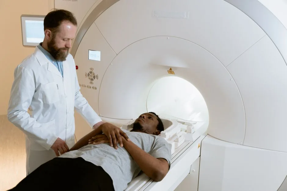
Unlike CT, MRI uses no ionizing radiation, making it safer for repeat imaging or for patients who need multiple followups. The scanner is also quieter than you might think (well, quiet compared to the hum of a hospital hallway), and most facilities offer headphones or music to ease any claustrophobia.
When CT still has a role
If a patient arrives at the emergency department with a suspected bleed, a quick noncontrast CT can rule out hemorrhage in under five minutes. Thats why you sometimes hear doctors say, Well do a CT first, then an MRI if needed. The key is that CT excels at spotting blood, while MRI shines at detecting tissue damage.
NonContrast MRI
Core sequences youll hear about
On a plain MRI for stroke, the radiologist focuses on a few essential sequences:
- DWI (DiffusionWeighted Imaging): Highlights water movement changes in brain cellsyour early warning sign.
- ADC (Apparent Diffusion Coefficient): Confirms that the bright spot on DWI truly represents restricted diffusion, not an artifact.
- FLAIR (FluidAttenuated Inversion Recovery): Shows how old or new the lesion might be.
- T2weighted images: Give a broader view of edema and surrounding tissue.
What a noncontrast MRI shows
When you get a plain scan, the radiologist is looking for:
- Hyperintense DWI lesionsthese are the hot spots that tell us a stroke has started.
- Loss of the dark signal on ADC mapssignifies true restricted diffusion.
- Peripheral FLAIR changeshelp determine if the stroke is hyperacute (minutes to hours) or evolving over days.
All of this happens without any injected dye, meaning theres no risk of contrastrelated side effects.
Limitations you should know
While noncontrast MRI is fantastic for spotting early ischemia, it cant always answer questions like:
- Are there subtle blood vessel abnormalities (aneurysms or arteriovenous malformations)?
- Is there a tiny bleed thats hidden on DWI?
- Has the bloodbrain barrier been compromised, suggesting a more complex pathology?
When any of those doubts arise, doctors may ask for a contrastenhanced study.
ContrastEnhanced MRI
Why add gadolinium?
Gadoliniumbased contrast agents act like a highlighter for the brains blood vessels and any areas where the bloodbrain barrier (BBB) is leaky. Doctors typically order a contrast MRI when they need to:
- Visualize vessel lumen and detect stenosis or occlusion (often through MRAMagnetic Resonance Angiography).
- Identify subacute hemorrhage that might be invisible on plain scans.
- Differentiate between tumor, infection, or inflammation that can mimic stroke symptoms.
What youll see after contrast
Postcontrast images can reveal:
- Enhancement of the infarcted coreindicating BBB breakdown.
- Clear delineation of arterial walls and any plaques.
- Contrast pooling in areas of active bleeding, helping clinicians decide if thrombolysis is still safe.
Risks, fasting, and preparation
Most people tolerate gadolinium without trouble, but there are a couple of things to keep in mind:
- Fasting: Youll usually be asked to skip food for 46 hours before the scan. The fast helps reduce the chance of nausea or vomiting after the contrast is injected.
- Kidney function: If you have chronic kidney disease, your doctor may run a quick blood test (creatinine) to ensure the contrast wont linger.
- Allergic reactions: Rare, but they can happen. If youve had a reaction before, let the technologist know right away.
Overall, the benefits of a contrastenhanced MRI in clarifying complex stroke cases far outweigh these modest risks.
Reading MRI Scans
Stepbystep quickcheck guide
Imagine youre sitting next to a radiologist, and they hand you the images. Heres a friendly cheatsheet you can keep in mind:
- Identify the sequence: Is it DWI, ADC, FLAIR, T1pre, or T1post (after contrast)?
- Look for diffusion restriction: Bright spot on DWI + dark on ADC = acute infarct.
- Assess enhancement: Any bright areas on postcontrast T1? That could mean BBB leakage or a hidden bleed.
- Check vessel patency: On MRA, see if the major arteries (MCA, ACA, PCA) are open or narrowed.
- Correlate clinically: Does what you see match the patients symptoms? If not, ask for a second opinion.
Comparison table: NonContrast vs. Contrast MRI
| Feature | NonContrast MRI | Contrast MRI |
|---|---|---|
| Sensitivity for early ischemia | ||
| Vessel lumen visualization | ||
| Detects subtle hemorrhage | Limited | |
| Patient preparation | No fasting needed | Fast 46 hrs + renal labs |
| Risks | None | Gadolinium reactions, renal concerns |
Realworld example
Last year, a friend of mine, Mark, went to the ER with sudden lefthand weakness. The initial CT was clean, so the neurologist ordered a noncontrast MRI. The DWI showed a tiny spot in the right MCA territoryearly ischemia. Because his symptoms lingered, they added a contrast MRA, which revealed a narrowing of the same artery. The extra detail guided the team to a targeted clotremoval procedure that saved a significant amount of brain tissue.
Decision Flowchart
How doctors choose the right study
Think of the imaging pathway like a chooseyourownadventure story:
- Suspected stroke Immediate noncontrast CT to rule out bleeding (fast, cheap).
- If CT negative or unclear Proceed to noncontrast MRI (DWI, ADC, FLAIR).
- Does DWI show restriction?
- Yes Treat as ischemic stroke. Consider adding contrast if you need vessel detail or suspect hemorrhagic transformation.
- No Look for alternative diagnoses (migraine, seizure, tumor). Contrast MRI often clarifies.
- Need vascular map? Order contrastenhanced MRA (or CTangiography if MRI unavailable).
This flow mirrors the recommendations from the American Heart Association/American Stroke Association, ensuring that each patient gets the most informative scan without unnecessary exposure.
Conclusion
Choosing between a plain MRI and a contrastenhanced MRI isnt just a technical decisionits a pivotal part of tailoring stroke care to each individuals needs. Noncontrast MRI gives you a rapid, safe snapshot of early brain injury, while adding gadolinium can illuminate hidden vessel problems, subtle bleeds, and bloodbrainbarrier leaks.
By understanding what each study shows, why fasting might be required, and how the images are read, youre better equipped to have an informed conversation with your neurologist or radiologist. Remember, the goal is always the same: to get the right picture as quickly as possible so that treatment can be started on time.
If you ever find yourself facing the MRI for stroke with or without contrast decision, ask your doctor about the specific reasons for each option, any preparation steps, and what the findings could mean for your treatment plan. Knowledge is power, and in stroke care, that power can literally save brain tissue.