Not medical advice — written by Ngoc Vietsmall, who is not a healthcare professional.
This article summarises publicly available medical information for general readers. It cannot account for your history, medication or symptoms, and it is not a diagnosis or a treatment plan. Any dose mentioned comes from published labelling or guidelines — only your own prescriber can decide what is right for you.
Always speak to a doctor or pharmacist before starting, stopping or changing any treatment. If symptoms are severe or getting worse, seek urgent care. Full disclaimer · How this is written
Ever glanced at a chest X-ray and wondered why the main pulmonary artery looks unusually wide, or why the tiny vessels at the lung edges seem to have vanished? If you are reviewing imaging for someone with portal hypertension and suspect a breathing-related issue, you might be looking at signs associated with portopulmonary hypertension radiology. In short, key indicators can include a dilated central artery, pruned peripheral vessels, and hints of right-heart enlargement. Recognizing these features can help guide discussions with healthcare providers about treatment plans.
Quick Answer Overview
What is portopulmonary hypertension on imaging?
Portopulmonary hypertension (POPH) may appear as an enlarged main pulmonary artery (often noted as greater than 30mm in medical literature), a noticeable thinning or loss of the fine peripheral vascular markings—what radiologists call pruning—and an enlarged right ventricle on both X-ray and CT. These three findings together are often described as the radiologic fingerprint of POPH.
Why should you care?
Because POPH sits at the crossroads of liver disease and heart-lung health. Detecting it early helps hepatologists decide whether a liver transplant is safe, and it guides pulmonary specialists in selecting targeted therapies. In other words, a sharp eye on the images can steer a whole cascade of care decisions.
Pathophysiology Quick Overview
How does portal hypertension lead to pulmonary arterial hypertension?
When the portal vein is backed up, substances that normally get cleared by the liver flood the circulation. Some of these vasoactive agents cause the pulmonary arteries to constrict and remodel, raising pressure on the right side of the heart. This cascade is what is referred to as portopulmonary hypertension.
Whats the difference between pulmonary arterial and venous hypertension on imaging?
Arterial hypertension, like POPH, shows a big central artery and pruned peripheral vessels. Venous hypertension, on the other hand, lights up the pulmonary veins, creates septal thickening and interstitial edema, and youll often spot Kerley B lines on a plain film. Understanding this distinction helps avoid mistaking one for the other.
Core Imaging Modalities
Chest X-Ray (CXR)
Classic CXR signs
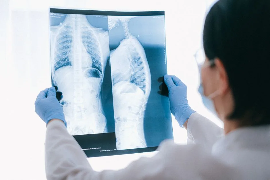
The most immediate clue is a widened main pulmonary artery—usually measured at the hilum on a posteroanterior view. Add to that a pruning pattern where the peripheral vasculature looks unusually sparse, and youve got a strong suspicion of POPH.
Measuring the main PA diameter
Radiologists measure the diameter at the level of the pulmonary artery bifurcation. Medical guidelines often cite cutoffs such as 29mm for women and 31mm for men as thresholds for further investigation. Only a qualified prescriber can determine the clinical significance of these measurements for an individual.
Pruning vs. other causes
Conditions like emphysema or chronic bronchitis can also thin out peripheral vessels but usually come with hyperinflated lungs and barrel-shaped chests. POPHs pruning is accompanied by a clearly enlarged central artery and right-heart enlargement.
CT Pulmonary Angiography (CTPA) & High-Resolution CT (HRCT)
CT findings specific to POPH
CTPA shows the same main-PA dilation (often greater than 30mm) plus a striking reduction in vessels under 2mm in diameter—a visual pruning thats easier to appreciate on cross-sectional images. Look for a right-ventricular/left-ventricular (RV/LV) ratio above 1.0; thats a red flag for pressure overload.
Dual-energy CT
Newer dual-energy techniques can color-code perfusion, helping you see areas of low blood flow that line up with the pruned vessels. According to a deep-learning model study, AI can even flag likely POPH cases from standard CT datasets with impressive accuracy.
Protocol checklist
When a CTPA is ordered for suspected POPH, protocols may specify contrast volumes around 80–100mL, a slice thickness of 1mm, and a bolus-tracking threshold of 150 HU in the main PA. These settings give you the detail needed to assess pruning and right-heart size.
MRI & Echocardiography (Supplementary)
When MRI adds value
If radiation is a concern or precise flow measurements are needed, cardiac MRI can quantify pulmonary artery flow, RV ejection fraction, and even detect subtle fibrosis.
Echo as a screening bridge
Transthoracic echocardiography is often the first step in many centers—look for a tricuspid regurgitation velocity greater than 3.4m/s. If its high, clinicians may pursue a CTPA or MRI for definitive anatomic detail.
Emerging AI & Deep-Learning Aids
AI-assisted detection
Recent AI tools trained on thousands of chest X-rays can automatically measure PA diameter and flag pruning, offering a second pair of eyes that never gets tired. While still under validation, these models are beginning to appear in radiology workstations.
Comparing Hypertension Types
| Feature | Portopulmonary Hypertension (POPH) | Pulmonary Arterial Hypertension (PAH) | Pulmonary Venous Hypertension (PVH) |
|---|---|---|---|
| Main PA size | >30mm, often >35mm | Enlarged but may be <30mm | Usually normal |
| Peripheral vessels | Pruned, <2mm | Similar pruning | Prominent, dilated veins |
| Lung interstitium | Clear, no edema | Clear | Septal thickening, Kerley B lines |
| Common comorbidities | Portal hypertension, liver cirrhosis | Connective-tissue disease, idiopathic | Left-heart disease, mitral valve disease |
| Key imaging differentiator | Pruning + liver-related signs (ascites, splenomegaly) | Isolated arterial changes | Venous dilation, pulmonary edema |
Practical Imaging Workflow
Suspicion & Reporting Checklist
Red-flag imaging signs
1 Main PA >30mm
2 Evident peripheral pruning on CXR or CT
3 RV/LV ratio >1.0 on CT/MRI
4 Associated liver disease on abdominal imaging
Structured report template
Radiology reports often start with a concise Impression line: Findings consistent with portopulmonary hypertension — dilated main PA (34mm), peripheral pruning, RV/LV ratio 1.2. This is followed by measurements, associated hepatic findings, and recommendation for further hemodynamic evaluation.
Multidisciplinary Communication
Talking to hepatology & cardiology
When POPH is flagged, communication with the liver team is essential. Notes might include PA diameter, pruning observations, and recommendations for right-heart catheterization before transplant evaluation. Clear, actionable language keeps everyone on the same page.
Follow-up imaging timeline
Follow-up schedules are determined by the treating physician. Baseline CT at diagnosis, a 6-month repeat if therapy changes, then yearly surveillance unless clinical status worsens are examples of how monitoring might be structured.
Pitfalls & Common Misinterpretations
Pruning vs. emphysema
Emphysema shows hyperinflated lungs and flattened diaphragms, while POPHs lungs stay relatively normal in volume. Use the PA diameter as the tiebreaker.
Obesity and PA size
In very obese patients, the PA can appear artificially wide on a single projection. Corroborate with CT or a lateral view before calling it POPH.
Patient-Centric Imaging Perspective
What patients can expect from imaging
Most of the work—CXR and CT—uses low to moderate radiation (a chest CT is roughly 2–3mSv). Contrast agents carry a small risk of allergic reaction, but prescreening and hydration usually keep things safe.
How imaging influences treatment
When images confirm POPH, the pulmonary team may consider endothelin-receptor antagonists or phosphodiesterase-5 inhibitors. Meanwhile, the liver transplant committee will weigh the severity of POPH against transplant benefits. In short, your radiologic read is a pivotal piece of the therapeutic puzzle.
Trusted patient resources
For anyone wanting plain-language explanations, the Pulmonary Hypertension Association offers clear brochures on what POPH means and why imaging matters.
Conclusion
Portopulmonary hypertension radiology hinges on three visual clues—an enlarged central pulmonary artery, pruning of peripheral vessels, and right-heart enlargement. Recognizing these signs early lets clinicians chart a safer, more effective treatment path, whether thats targeted medication or a carefully timed liver transplant. Use the checklist, stay in touch with the multidisciplinary team, and dont shy away from leveraging new AI tools that can catch subtle patterns you might miss. By keeping the patients whole picture in mind, you turn every scan into a step toward better health.