Not medical advice — written by Ngoc Vietsmall, who is not a healthcare professional.
This article summarises publicly available medical information for general readers. It cannot account for your history, medication or symptoms, and it is not a diagnosis or a treatment plan. Any dose mentioned comes from published labelling or guidelines — only your own prescriber can decide what is right for you.
Always speak to a doctor or pharmacist before starting, stopping or changing any treatment. If symptoms are severe or getting worse, seek urgent care. Full disclaimer · How this is written
Short answer: a CT scan can show changes that suggest a stroke has already happened (including silent ones that caused no noticeable symptoms), but it cannot predict a future stroke with certainty. Knowing what a CT can and cannot show can help you and a healthcare professional make more informed decisions when time matters.
Why does this matter to you? Imagine you are at home, a friend suddenly slurs their words, and you are trying to understand what to do. The right imaging test can be the difference between “we call emergency services now” and “we wait and watch.” Let’s cut through the jargon together and get clear on what the scan actually does.
Quick Answer
Can a CT scan detect a stroke before it happens?
No. A CT (computed tomography) captures what is already inside the brain at the moment of the scan. If there is bleeding, tissue swelling, or an old scar, the CT may show it. But it cannot read the future or see a clot that has not yet caused injury.
Why CT isnt a crystalball
Think of a CT like a snapshot. It freezes the current scene—bright white bone, gray brain tissue, and any dark areas that may represent bleeding. It cannot sense the early activity of a clot forming in a vessel that looks normal right now. That is why healthcare professionals often combine imaging with clinical signs, such as sudden facial droop, and risk-factor checks such as blood pressure and heart rhythm to decide what comes next.
Imaging the present vs. predicting the future
- CT can show: acute bleeding, large-area swelling, calcifications, and old infarcts that may appear as silent scars.
- CT cannot show: plaque that might rupture tomorrow, microvascular disease, or a clot that has not yet caused tissue injury.
- What that means for you: a normal CT does not guarantee you will not have a stroke later; it simply means no visible injury was seen at the time of the scan.
CT Scan Basics
How quickly does a stroke show up on a CT?
In the first minutes, sometimes called the hyperacute window of 0 to 3 hours, CT is often excellent at detecting hemorrhage, or bleeding. Early ischemic changes—like subtle loss of gray-white differentiation—can be faint, but radiologists are trained to look for them. After about 6 to 12 hours, the scan may show more obvious changes in the affected area.
Detecting an existing stroke vs. an emerging one
If a clot is already reducing blood flow, the CT may begin to show changes in the affected tissue. The earlier the scan, the subtler the signs may be, which is why emergency departments often perform the scan quickly after symptoms begin.
Timing windows
| Phase | Typical CT Findings |
|---|---|
| Hyperacute (0 to 3 hours) | Often clear for hemorrhage; possible subtle loss of gray-white contrast. |
| Early (3 to 24 hours) | Visible dark area may appear where tissue is injured; swelling may begin. |
| Subacute (1 to 7 days) | Well-defined low-density region may be seen; swelling may peak. |
| Chronic (more than 2 weeks) | Permanent low-density scar may remain; surrounding tissue may shrink. |
CT vs. MRI for early detection
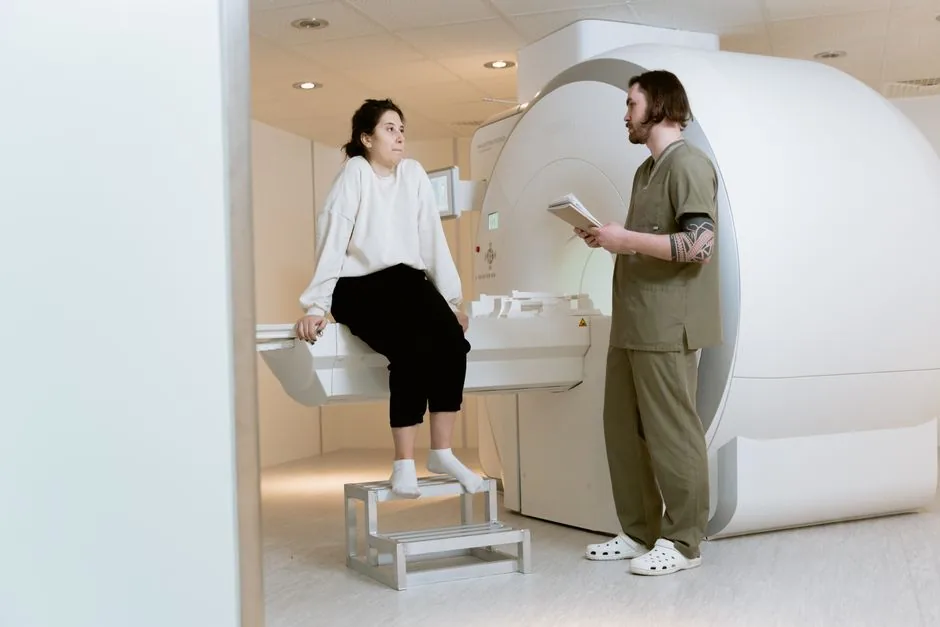
Both CT and MRI can help detect changes from a stroke, but they have different strengths. CT is fast, widely available, and often used first to check for bleeding. MRI, especially diffusion-weighted imaging, can show small ischemic changes very early but takes longer and may not always be available in every emergency department.
CT vs. MRI Comparison
| Aspect | CT Scan | MRI |
|---|---|---|
| Speed | About 5 to 10 minutes | About 15 to 30 minutes |
| Availability | Most hospitals | Often limited to larger centers |
| Radiation | Small amount | None |
| Hemorrhage detection | Excellent | Very good |
| Early ischemia detection | Moderate | High sensitivity |
MiniStroke Imaging
Does a ministroke show up on a CT scan?
Often, no. Mini-strokes—technically called transient ischemic attacks, or TIAs—are brief interruptions of blood flow. By the time the CT is done, the brain may look normal because the episode may not leave changes that a CT can detect.
When MRI is preferred for TIA detection
MRI with diffusion-weighted imaging can sometimes detect signs of a recent TIA that a CT may miss. That is one reason a healthcare professional may recommend MRI when a TIA is suspected but the CT does not show a clear cause.
Realworld case example
Consider a hypothetical example: a 58-year-old accountant felt sudden right-sided weakness that cleared within 10 minutes. A CT scan showed no clear abnormality. A later MRI showed a small area consistent with a recent transient ischemic event. A healthcare professional may then discuss options such as aspirin and lifestyle changes to help lower future risk.
MRI vs. CT: Which Is Better?
Can an MRI detect a stroke before it happens?
Like CT, MRI visualizes what is already present. It can be very good at showing early changes, but it cannot forecast a future clot. The phrase “before it happens” is a myth that many online sources repeat.
How long will a stroke show up on an MRI?
Diffusion-weighted images can highlight acute injury within minutes and may stay bright for about 10 to 14 days. After that, the signal may fade, and other MRI sequences, such as FLAIR and T2, can help show the evolving area.
Pros & Cons Table
| Factor | CT Scan | MRI |
|---|---|---|
| Speed | Fast, about 5 to 10 minutes | Slower, about 15 to 30 minutes |
| Cost | Generally lower | Often higher |
| Radiation | Small amount | None |
| Hemorrhage detection | Excellent | Very good |
| Early ischemia detection | Moderate | High sensitivity |
| Availability | Widespread | Often limited to larger centers |
Beyond Imaging
How to test for stroke at home
While nothing replaces a professional evaluation, you can stay aware of possible warning signs:
- Check your blood pressure regularly, because high readings can be an important risk factor.
- Use the FAST test: Face drooping, Arm weakness, Speech trouble, Time to call emergency services.
- Consider online risk calculators, such as the tool from the American Heart Association.
Preventive measures & early warning signs
Some lifestyle changes may help lower stroke risk over time. Think of them as maintenance for the brain’s blood vessels:
- Avoid smoking, because smoking can contribute to plaque buildup over time.
- Talk with a healthcare professional about cholesterol, diet, exercise, or medication if needed.
- Discuss heart rhythm concerns, because atrial fibrillation can raise clot risk.
- Regular physical activity, such as brisk walking, may support vascular health.
Reader checklist
- Know your blood pressure.
- Recognize FAST warning signs.
- Have a healthcare professional review your heart rhythm.
- Schedule regular checkups if you have diabetes or high cholesterol.
Interpretation Risks
Benefits of early CT detection
When a stroke is suspected, a CT can help the medical team quickly decide whether certain time-sensitive treatments, such as clot-dissolving medication or mechanical thrombectomy, may be appropriate. A CT that shows no bleeding can be one factor in clearing the way for those treatments, but eligibility is determined by a treating clinician.
Risks &limitations
CT involves a small amount of radiation—some compare it to a few days of natural background exposure—but it is still something to consider, especially with repeated scans. Also, a normal CT in the first hour may not show a small area of early ischemia, so symptoms should still be evaluated carefully.
Expert comment
A normal CT should not be used alone to rule out a stroke. Clinical assessment remains important; imaging is one tool, not a replacement for a full evaluation.
Treatment Pathways
What is the best treatment for stroke?
The best plan depends on the type of stroke and the person’s situation:
- Ischemic stroke, the most common type: clot-dissolving medication may be an option within a time window described on the official label, or mechanical thrombectomy may be considered if a large vessel is blocked. Only the treating clinician can determine whether these options are appropriate.
- Hemorrhagic stroke: blood pressure management, possible surgery, and careful monitoring may be used.
How imaging guides treatment choice
A CT that shows bleeding can shift the plan away from clot-dissolving medication and toward possible surgical or supportive care. A CT or CT angiography that shows a clot in a large vessel may prompt consideration of mechanical thrombectomy. Imaging is one factor the care team uses to evaluate possible options.
Decisionmaking flowchart (for illustration)
- Sudden symptoms, such as face drooping, arm weakness, or speech difficulty, may prompt a FAST check and a call to emergency services.
- In many emergency departments, a noncontrast CT is often one of the first imaging tests.
- If the CT shows bleeding, the care team may consider a hemorrhagic pathway.
- If the CT does not show bleeding, additional imaging such as CT angiography may be used to look for a large-vessel blockage.
- If a large-vessel blockage is present, mechanical thrombectomy may be considered, sometimes along with clot-dissolving medication if the person is eligible.
- If no large-vessel blockage is found, the team may focus on medical support, monitoring, and secondary prevention.
Putting It All Together
Let’s recap the essentials in a friendly, bite-size way:
- A CT scan can show changes that suggest a stroke has already happened—old, silent, or acute—but it cannot predict a future event.
- For emergency symptoms, CT is often the fastest way to check for bleeding and help a care team decide whether clot-dissolving therapy may be safe.
- Mini-strokes often look normal on CT; MRI may be recommended if a TIA is suspected.
- Both CT and MRI have pros and cons; the choice depends on speed, availability, and what the healthcare professional needs to see.
- Do not rely on imaging alone; know your risk factors, recognize FAST warning signs, and keep up with preventive care.
- When a stroke is confirmed, imaging can help shape the treatment plan—whether that involves clot-dissolving medication, thrombectomy, or surgical intervention.
Remember, imaging technology can be a helpful tool, but recognizing warning signs and acting quickly is important. If you or someone else experiences possible stroke warning signs, call emergency services right away—do not wait for a scan to confirm what you are seeing.
What questions do you have about CT or MRI after possible stroke symptoms? Share your thoughts or questions in the comments below; your story might help someone else feel a little less scared.