Not medical advice — written by Ngoc Vietsmall, who is not a healthcare professional.
This article summarises publicly available medical information for general readers. It cannot account for your history, medication or symptoms, and it is not a diagnosis or a treatment plan. Any dose mentioned comes from published labelling or guidelines — only your own prescriber can decide what is right for you.
Always speak to a doctor or pharmacist before starting, stopping or changing any treatment. If symptoms are severe or getting worse, seek urgent care. Full disclaimer · How this is written
Ever glance at your forearm or leg and wonder, What are these tiny white spots? You're not alone—most of us have stared at a puzzling patch and felt a mix of curiosity and a dash of worry. In the next few minutes we'll walk through what those spots might be, why they can appear, when they could be a signal to see a clinician, and how they may be managed or prevented. Think of this as a friendly chat over coffee, with clear photos, real-life stories, and practical tips right at your fingertips.
Quick Visual Guide
Before we dive into the science, let's get a quick snapshot of what you might be looking at. White spots come in several shapes, sizes, and textures. Knowing the basics can help you describe what you notice when you talk with a clinician.
Typical Appearances
- Milia – tiny, firm, pearl-white bumps, usually 1 to 2 mm, most often on the cheeks.
- Vitiligo patches – irregular, smooth, milky-white areas that can range from a few millimeters to large sections.
- Fungal-related spots – slightly scaly, often on the legs or torso, with a pale-white center and a pinkish border.
- Sun-damage hypopigmentation – faded, flat spots where pigment may have been reduced by UV exposure.
- Vitamin-deficiency spots – diffuse, fine, peppercorn-like speckles, especially on the legs in B12 deficiency.
If you take a quick photo of the spot, noting its size, texture, and location can be useful for the DIY Photo Check later on.
Common Medical Causes
| Possible Cause | Possible Reason | What May Be Seen |
|---|---|---|
| Sun-induced hypopigmentation | UV exposure may reduce pigment in some areas. | Flat, faded areas on sun-exposed skin. |
| Milia | Keratin may become trapped under the outer layer. | Hard, tiny pearl-white bumps, mainly on cheeks. |
| Vitiligo | An autoimmune process can affect pigment-producing cells. | Sharp, irregular patches that may spread. |
| Tinea versicolor (fungal) | A type of yeast may temporarily alter skin color. | Scaly, slightly pink-white spots, often on trunk. |
| Pityriasis alba | Mild eczema may leave a pale patch after healing. | Soft, dry, pale areas on the face of children. |
| Post-inflammatory hypopigmentation | Healing from a rash, burn, or injury may reduce pigment for a time. | Faded area matching the shape of the original inflammation. |
| B12 vitamin deficiency | Low vitamin B12 may affect pigment production. | Fine, pepper-like pale specks, typically on legs. |
| Calcium / Vitamin D deficiency | May be associated with tiny, pale spots. | Small, pinpoint spots, often on arms. |
| Skin cancer (basal cell, melanoma) | Some skin cancers may appear as a pale or scar-like area. | Asymmetric, growing, possibly ulcerated spots. |
| Lichen sclerosus | Chronic inflammation may cause thin, pale plaques. | White, fragile patches, usually in the genital area. |
| Pityriasis lichenoides chronica | Small papules may fade to white. | Pinpoint flat white lesions on trunk. |
| Drug-induced hypopigmentation | Some medications may affect pigment production. | Uniform fading where medication was applied. |
This table lists some possibilities, but it is not a diagnostic tool. If you recognize a pattern from the table, that can be a useful starting point for a conversation with a clinician.
When to Worry
Most white spots are harmless, but a few features may warrant prompt medical attention. Here are some signs to discuss with a clinician.
Red-Flag Signs
- Asymmetry – one side looks different from the other.
- Irregular, jagged borders.
- Rapid growth or change in color.
- Bleeding, crusting, or ulceration.
- Any spot that feels painful or itchy after a short period.
Usually Less Concerning vs. Worth a Medical Evaluation
| Feature | Usually less concerning | Worth a medical evaluation |
|---|---|---|
| Shape | Round, smooth, uniform | Irregular, asymmetric |
| Border | Well-defined, clean | Blurry or jagged |
| Change Over Time | Stable or slowly evolving | Rapid enlargement or new color |
| Sensation | Usually none | Pain, itching, bleeding |
If any of those red-flag characteristics match a spot you have photographed, consider bringing the picture to a dermatologist. A visit can help clarify what is going on and offer peace of mind.
Nutrient Deficiencies Explained
Sometimes the appearance of pale spots may be linked to nutrition. Low levels of certain nutrients can be associated with tiny, pale speckles that look similar to milia or early vitiligo.
B12 Deficiency and White Spots
Vitamin B12 is important for several body processes, including those that involve skin color. When levels are low, some people notice tiny pale spots on the skin, especially on the lower legs. These spots are often uniform and may occur alongside fatigue, tingling sensations, or mood changes.
Other Deficiencies
- Vitamin A – dry or rough patches may appear as lighter areas.
- Vitamin D – low levels sometimes coincide with lighter areas.
- Calcium – low levels may be associated with small pale specks.
How Testing and Nutrition May Help
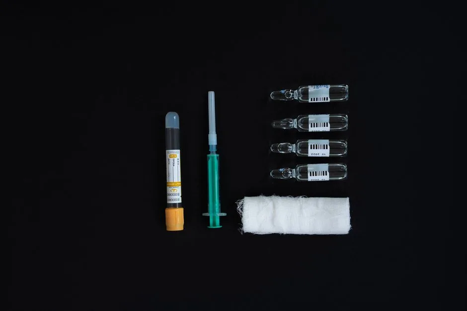
If you are concerned, you can talk with a clinician about whether a blood test may be helpful. These panels sometimes include vitamin B12, vitamin D, calcium, and a basic metabolic screen. If any values are low, a clinician can suggest whether a supplement or dietary change is appropriate. A balanced diet with leafy greens, fortified cereals, fish, and dairy may also support overall skin health.
Fungal Infections Guide
Fungal organisms can overgrow on warm, moist skin. When that happens, pale, sometimes scaly patches can appear and may look similar to other conditions.
Tinea Versicolor
Tinea versicolor may appear as pale or slightly pink patches, often on the chest, back, and upper arms.
Treatment Options
- Topical antifungals – Non-prescription products containing selenium sulfide are sometimes used for tinea versicolor. Label instructions should be followed, and a clinician can help decide whether this is appropriate.
- Oral antifungals – For widespread cases, a clinician may prescribe a short course of an oral antifungal such as fluconazole (Mayo Clinic).
- Skin care – Some people find it helps to keep the area dry, wear breathable fabrics, and change out of sweaty clothes promptly.
Many fungal-related spots respond well to treatment, though they can recur.
DIY Photo Check
Your phone camera can be a useful record-keeping tool. These steps may help you gather details before a dermatologist appointment.
Step-by-Step Visual Checklist
- Lighting – Natural daylight is often best if available; a harsh flash may make details harder to see.
- Focus – A close, stable camera position can help show edges clearly.
- Size – Measuring with a ruler or credit-card tip can help document the diameter.
- Texture – Note whether the spot is smooth, scaly, or raised.
- Location – Keeping a record of the body location can be useful.
- Number – Note whether it is a single spot or multiple, and whether they are grouped.
Once you have this information, you can use the Typical Appearances section as a general reference, but it cannot tell you which condition you have. If you notice any red-flag features, consider uploading the photo to a clinician's portal or bringing a printed copy to an appointment.
Treatment Overview Guide
Once a clinician has helped identify the likely cause, it can be useful to understand the range of treatment approaches. Options may include simple skin care adjustments or professional procedures.
Topical Options
- Hydroquinone – a prescription skin-lightening ingredient sometimes used under specialist supervision for certain kinds of discoloration.
- Retinoids (such as tretinoin) – may support skin turnover and be helpful for some cases of milia under clinician supervision.
- Barrier-repair creams (ceramides, hyaluronic acid) – may support the skin barrier in post-inflammatory hypopigmentation.
Oral Therapies
- Vitamin B12 supplements may be considered when a deficiency is confirmed by a healthcare professional.
- Antifungal tablets are sometimes prescribed for widespread tinea versicolor.
- Short courses of systemic steroids may be prescribed by a specialist for severe vitiligo flares.
Procedural Interventions
- Laser therapy (excimer, fractional CO) – may support repigmentation in some people with vitiligo.
- Phototherapy (NB-UVB) – may be an option for some people with widespread vitiligo or psoriasis-related pigment loss.
- Minor surgical removal – may be an option for persistent milia when other approaches have not helped.
Every treatment carries pros and cons. For example, laser sessions may require multiple visits and a recovery period, while topical creams may need daily commitment. A clinician can help you weigh the options based on your specific situation.
Prevention & Lifestyle Tips
Daily habits may support skin health and help you feel more comfortable. Here are some practical steps many people find helpful.
- Sun protection – A broad-spectrum sunscreen with SPF 30 or higher is a common choice for sun-exposed skin, even on cloudy days.
- Gentle cleansing – You may find it helpful to avoid harsh soaps that strip natural oils.
- Balanced nutrition – A diet that includes B-vitamin-rich foods (eggs, fish, fortified cereals) and vitamin D sources (sunlight, fortified milk) may support skin health.
- Regular skin checks – A monthly self-check can help you notice any new or changing spots; a clinician can show you what to look for.
- Stay dry – Changing out of sweaty clothes promptly, especially after exercise, may help reduce moisture buildup.
These habits may support skin health, but they are not a guaranteed way to prevent or treat any specific condition.
Real World Stories
Stories can make the science feel real. Here are two brief accounts that illustrate how a medical evaluation can help clarify what is happening.
Case Study A – Milia on the Cheeks
Emily, a 28-year-old graphic designer, noticed a cluster of pearl-white bumps on her cheeks after a month of intensive makeup use. She photographed them and initially tried over-the-counter exfoliants, which she felt made them worse. A quick visit to her dermatologist suggested milia, which can be associated with heavy occlusive creams. After her clinician recommended a non-comedogenic moisturizer and prescribed a gentle retinoid, her bumps improved over six weeks. Emily's experience shows how a product change may be helpful, but results can vary.
Case Study B – B12 Deficiency on the Legs
Mark, a 52-year-old accountant, started noticing snowflake-like speckles on his shins during his morning jogs. He thought they were age spots, but the pattern reminded him of descriptions of B12 deficiency. Blood tests confirmed low cobalamin. After his clinician recommended B12 injections and dietary adjustments, the speckles improved dramatically. Mark now shares his journey on a health forum, encouraging others to consider medical screening rather than assuming the spots are harmless.
Both stories highlight how a clear photo, a bit of curiosity, and professional guidance can help turn confusion into confidence.
Conclusion
White spots on the skin can be associated with many things, from milia to nutrient levels to changes that need a closer look. Using a visual checklist, learning about common possibilities, and knowing when to seek professional help can give you a solid starting point. A clear photo and guidance from a clinician can help you understand what you are seeing and what options may be available. Stay curious, stay protected, and do not hesitate to share your own spot story with a trusted friend or medical professional. Your skin's health is a journey, and you are now equipped to travel it with confidence.