Not medical advice — written by Ngoc Vietsmall, who is not a healthcare professional.
This article summarises publicly available medical information for general readers. It cannot account for your history, medication or symptoms, and it is not a diagnosis or a treatment plan. Any dose mentioned comes from published labelling or guidelines — only your own prescriber can decide what is right for you.
Always speak to a doctor or pharmacist before starting, stopping or changing any treatment. If symptoms are severe or getting worse, seek urgent care. Full disclaimer · How this is written
At first, I thought it was nothingjust a little haze when I walked into a brightly lit room. Then I started seeing halos around lights, reading menus felt like looking through frosted glass, and I wondered, What does vision really look like when cataracts set in? The short answer: blurry, hazy, and a bit glarefilled. Below youll find real photos that show exactly how cataracts change what you see, plus sidebyside comparisons of normal eyes, earlystage cloudiness, and the crisp clarity after surgery. Lets dive in together.
Why Cataract Photos Matter
What does vision look like with cataracts?
Imagine trying to watch a movie on a screen thats halfcovered with a sheer curtain. Thats the everyday reality for many people with cataracts. The images youll see here illustrate three core distortions:
- Blur: Objects lose sharp edges, making text hard to read.
- Glare and halos: Light sources explode into rings, especially at night.
- Colour washout: Bright yellows and blues become muted, giving the world a pastel tone.
These visual changes arent just inconvenientthey can affect safety, like driving after dark. Seeing the photos helps you recognize the signs before they impact daily life.
Early cataract stages pictures
In the first few years, cataracts develop slowly. Below is a gallery of early cataract stages pictures that show a faint cloudiness right in the centre of the lens (the nuclear region). Most people describe this stage as the world feels a little foggy, but I can still manage. The images are sourced from ophthalmology clinics and illustrate exactly what to look for when you next notice a subtle shift in vision.
Normal eye and cataract eye images
Sidebyside normal eye and cataract eye images make the contrast crystal clear. In a normal eye, you see crisp lines and vivid colours. In a cataractaffected eye, the same scene looks muted and smeared. This visual comparison is the quickest way to spot whether your vision has truly changed or if youre just tired.
Cataract Types Visuals
Nuclear, cortical, and posteriorsubcapsular
There are three main cataract types, each with a distinctive look on photos:
- Nuclear: A dense, yellowbrown cloud that starts in the centre of the lens.
- Cortical: Spokes radiating outward, giving a starburst appearance.
- Posteriorsubcapsular (PSC): A small, dense spot directly behind the lens, often the worst culprit behind nighttime glare.
When you glance at types of cataract images, youll notice the PSC form creates a dark pupillike shadow that moves as you look around. Knowing which pattern youre seeing can guide your doctor toward the most appropriate treatment plan.
What does blurry vision from cataracts look like?
Blurry vision isnt just out of focus. Its a layered distortion: the whole scene is softened, but bright lights create bright circles that bleed into surrounding colours. The photo below (illustrative) shows a street sign thats legible only after the glare fadessomething many patients describe as reading through a veil.
Surgery Photo Comparisons
Pictures of vision before and after cataract surgery
One of the most hopeful moments is flipping from a hazy picture to a crystalclear one after surgery. Heres a sidebyside set from a reputable eye centre that demonstrates the transformation. The after image shows dramatically improved contrast, colour fidelity, and depth perception.
According to a study from the Mayo Clinic, most patients experience a 2 to 3line gain on the Snellen chartessentially, they can read smaller print than before the operation.
What does a cataract look like when removed?
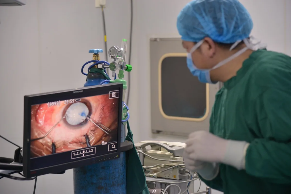
During phacoemulsification, the cloudy lens is broken up with ultrasonic waves and gently aspirated. Intraoperative photos reveal a clear, fluidfilled capsule once the cataract is gone, followed by the insertion of a thin, clear artificial lens (IOL). Seeing these images demystifies the surgery and can ease anxiety for anyone hesitant about the procedure.
SelfScreening Using Photos
Spotting warning signs in everyday vision
Do you notice any of these everyday clues?
- Halos around headlights or streetlights at night?
- Colors looking washed out, especially in bright sunlight?
- Frequent need to squint or hold reading material farther away?
Match your experience with the photos in this article. If you see a strong similarity, its time to schedule an eye exam. Remember, selfscreening is a friendly first stepnot a substitute for professional diagnosis.
Safe selfassessment tools
Some reputable apps let you simulate cataract vision on your smartphone, helping you compare what you see now versus a clouded view. Look for apps that are FDAcleared and that provide clear disclaimers that the simulation is educational only. If the simulated image feels familiar, thats a solid cue to book a comprehensive eye exam.
Common Cataract FAQs
| Question (keyword) | Quick Answer |
|---|---|
| What does blurry vision from cataracts look like? | It appears as a uniform fog with brightlight halos and reduced contrast. |
| Can I see a difference between a normal eye and a cataract eye in photos? | Yesnormal eyes show crisp, vibrant images; cataract eyes look hazy and glarefilled. |
| How many types of cataract images exist? | Three main types: nuclear, cortical, and posteriorsubcapsular, each with a distinct pattern. |
| Are beforeandafter cataract surgery pictures reliable? | When sourced from accredited clinics, they accurately reflect typical visual improvement. |
| Where can I find a safe VR experience of cataract vision? | Search for cataract virtual reality experience on reputable medical YouTube channels. |
Benefits and Risks
Benefits of using vision photos
Photos give you a visual language to describe whats happening inside your eye. They help you:
- Recognise early symptoms before they worsen.
- Communicate clearly with your eye doctor.
- Feel less anxious about surgery by seeing real outcomes.
Potential risks
Seeing dramatic photos can be unsettling, especially if youre already worried about your eyesight. Its easy to misinterpret a photo and assume you have cataracts when another condition is responsible. Thats why its crucial to pair selfscreening with a professional exam and not rely solely on images.
How clinicians mitigate risk
Eye specialists use these photos as teaching tools during consultations. They walk you through each picture, explain why it matchesor doesnt matchyour symptoms, and then perform a dilated exam for a definitive diagnosis. This collaborative approach balances the educational power of photos with the safety of clinical expertise.
Conclusion
Seeing real photos of vision with cataracts can turn a vague feeling of somethings off into a concrete understanding of whats happening inside your eye. Whether youre looking at earlystage cloudiness, comparing normal and cataract eye images, or marveling at the dramatic transformation after surgery, these visuals empower you to actwhether that means scheduling a checkup, learning about the different cataract types, or simply feeling less anxious about the upcoming procedure.
Remember, the images are a guide, not a diagnosis. If any of the pictures resonate with your experience, reach out to an eyecare professional you trust. Your vision is priceless, and a clear pictureboth literally and figurativelystarts with the right information and the right doctor.