Not medical advice — written by Ngoc Vietsmall, who is not a healthcare professional.
This article summarises publicly available medical information for general readers. It cannot account for your history, medication or symptoms, and it is not a diagnosis or a treatment plan. Any dose mentioned comes from published labelling or guidelines — only your own prescriber can decide what is right for you.
Always speak to a doctor or pharmacist before starting, stopping or changing any treatment. If symptoms are severe or getting worse, seek urgent care. Full disclaimer · How this is written
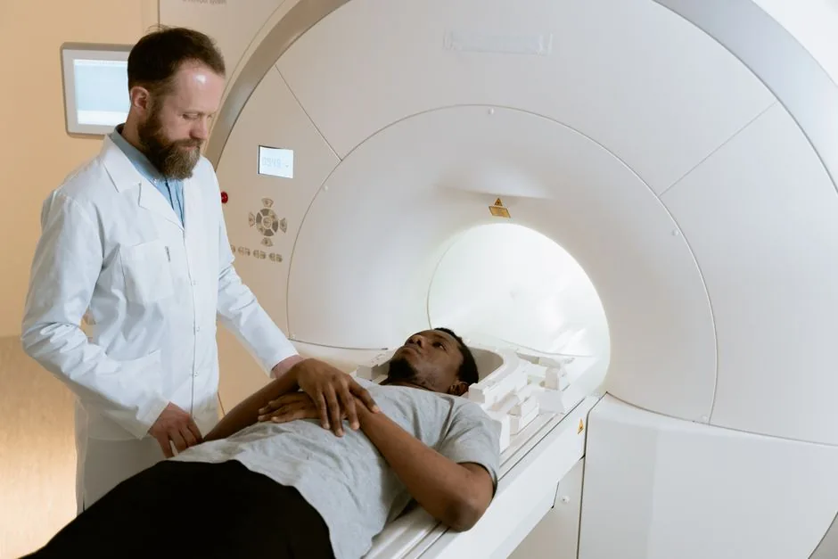
If you’re staring at a hospital brochure and wonder whether an MRI will actually show a stroke, you’re not alone. In the next few minutes you’ll get the straight-to-the-point answers about mri scan for stroke, how it stacks up against a CT, how long the findings may linger, and what it can mean for treatment. Let’s dive in as if we were chatting over a cup of coffee.
Quick Overview
Think of imaging like a flashlight in a dark room. An MRI shines a bright, detailed beam that can reveal small changes in brain tissue within the first hours, while a CT is more like a quick sweep that catches larger problems but sometimes misses subtle changes.
What MRI Shows
An MRI can reveal blood vessel blockages, bleeding, tissue changes, and even the penumbra — the area of brain that may still be at risk but might be salvageable. This is why healthcare teams often find it useful for ischemic stroke evaluation.
When Its Ordered
Most emergency departments start with a CT because it is fast. If the CT is inconclusive or the care team needs a clearer map of the vessels, they may order an mri or ct scan for stroke next.
Key Benefits
- No ionizing radiation, which may be a consideration for repeat scans.
- DiffusionWeighted Imaging (DWI) can sometimes detect changes within the first hours after symptoms begin.
- Excellent at distinguishing between hemorrhagic and ischemic events.
Key Risks
- Not suitable for people with certain metal implants or severe claustrophobia.
- Gadolinium contrast (used in mri for stroke with or without contrast) can be a concern for those with kidney problems.
MRI vs CT
Core Differences
Both scans can be helpful, but they have different strengths. Below is a quick side-by-side comparison that you can bookmark.
| Feature | CT Scan | MRI (with/without contrast) |
|---|---|---|
| Detects ischemic stroke | May show changes within hours but can miss very early changes | Can show changes within the first hours with DWI |
| Detects hemorrhage | Immediate | Immediate |
| Radiation exposure | Yes | No |
| Typical acquisition time | Usually shorter | Usually longer |
| Cost | May be lower, but varies widely | May be higher, but varies widely |
According to Mayo Clinic, early diffusion changes on MRI can help inform time-sensitive treatment decisions.
When to Choose Which
If you are in the ER with sudden facial droop, the first scan may be a CT because it is fast and can catch life-threatening bleeds quickly. After that, if the care team suspects an ischemic event, they may request an MRI to gather more detail for the treatment plan.
How MRI Detects Stroke
Important Sequences
Modern MRI machines run several sequences, each highlighting a different piece of the puzzle:
- DWI (DiffusionWeighted Imaging) — the early alarm bell; bright spots can mean water movement is restricted, which may suggest a fresh infarction.
- FLAIR (FluidAttenuated Inversion Recovery) — can show how old a lesion may be; the longer it has been present, the brighter the area may appear.
- MRA (Magnetic Resonance Angiography) — maps the arteries, sometimes with gadolinium.
- SWI (SusceptibilityWeighted Imaging) — can reveal microbleeds and calcium deposits.
Minicase Story
Last year a close friend experienced stroke-like symptoms. The CT looked normal, but the DWI on her MRI showed a small bright area in the left parietal lobe. That finding gave the care team additional information to decide on next steps. It was a reminder of how MRI can provide useful detail, even when symptoms are brief.
Visibility Timeline
How Long Will a Stroke Show Up on an MRI?
Answering the exact question many people search for:
- Acute phase — DWI may show changes very early, sometimes within the first hours, and can remain bright for several days.
- Subacute phase — FLAIR may become more obvious over the following days, and the lesion may be visible on both DWI and FLAIR.
- Chronic phase — Changes may remain visible long term, and imaging can sometimes help distinguish older areas from newer ones.
How Long Will a Stroke Show Up on a CT Scan?
CT can be slower to show early ischemic changes. In some cases, these changes may not be visible in the first few hours. Once an area becomes apparent, it can remain visible for some time on follow-up scans.
Reading an MRI Brain for Stroke
What Radiologists Look For
If you ever glance at an MRI report, these are the key phrases you may see:
- Diffusion restriction — bright on DWI, dark on ADC maps — may suggest an acute infarct.
- Hemorrhage on SWI — dark blooming spots — may suggest bleeding.
- Vessel occlusion on MRA — missing flow signal — may suggest a possible clot.
- Edema on T2/FLAIR — swelling around the lesion.
Tips for Patients
- You can ask the radiologist to explain whether the report describes core versus penumbra, which can give a sense of already injured tissue versus tissue that may still be at risk.
- Bring any previous scans; comparison can help track changes over time.
- If contrast was used, you can ask about any enhancement mentioned in the report, as it may point to a breakdown of the blood-brain barrier.
MRI With or Without Contrast
When Contrast Helps
Gadolinium-based contrast agents enhance the visibility of blood vessels and can highlight areas where the blood-brain barrier is leaky, which may be useful for:
- Detailed MRA studies to pinpoint a clot’s exact location.
- Identifying tumorous lesions that might mimic stroke.
- Assessing post-stroke inflammation.
Risks of Gadolinium
Most people tolerate gadolinium well, but if you have severe kidney disease, the risk of Nephrogenic Systemic Fibrosis (NSF) may be higher. Let your healthcare team know about kidney issues or previous allergic reactions.
Risks, Benefits & Practical Tips
Benefits You’ll Appreciate
- Early, accurate diagnosis — may help inform time-sensitive treatment decisions.
- No radiation — may be a consideration for younger people or repeated imaging.
- Detailed vessel mapping — can help plan surgical or endovascular procedures.
Potential Drawbacks
- Claustrophobia — the tunnel can feel tight, but many centers offer ways to help with comfort.
- Metal implants — pacemakers, cochlear implants, or certain aneurysm clips may be incompatible.
- Contrast allergies — uncommon, but mention any known allergies to your healthcare team.
Preparation Checklist
- Ask whether you need to remove jewelry, watches, or hairpins before the scan.
- Ask about wearing comfortable, metal-free clothing.
- If contrast is planned, confirm any eating or drinking instructions with the imaging center or your healthcare provider, especially if you have diabetes.
- Bring a current medication list, since some medications can affect kidney function.
- Mention any implants, metal fragments, or possible pregnancy concerns to the technologist.
Emergency Action Plan
When to Call 911
If you notice any of these sudden symptoms, treat them as a possible medical emergency and seek help fast:
- Sudden numbness or weakness, especially on one side.
- Difficulty speaking or understanding speech.
- Blurred or loss of vision in one eye.
- Severe, unusual headache.
- Loss of balance or coordination.
Remember, an MRI is a diagnostic tool — it is not a substitute for calling emergency services. Quick medical attention, followed by appropriate imaging (often CT first, then MRI), can improve the chance of a better outcome.
Conclusion
In a nutshell, an mri scan for stroke can be a very sensitive way to evaluate possible ischemic changes early, especially with DWI, and it can provide a detailed map of the brain’s blood vessels when contrast is used. It may be especially helpful for spotting small or subtle events, but CT remains a rapid first imaging choice in many emergency settings. Consider the benefits (no radiation, precise tissue detail) against the risks (metal implants, contrast concerns) and discuss any worries with a healthcare provider.
Feeling a bit overwhelmed? Grab a printable cheatsheet of the key points, or reach out to a healthcare professional to walk through your MRI report together. You deserve clear answers and a confident path forward.