Not medical advice — written by Ngoc Vietsmall, who is not a healthcare professional.
This article summarises publicly available medical information for general readers. It cannot account for your history, medication or symptoms, and it is not a diagnosis or a treatment plan. Any dose mentioned comes from published labelling or guidelines — only your own prescriber can decide what is right for you.
Always speak to a doctor or pharmacist before starting, stopping or changing any treatment. If symptoms are severe or getting worse, seek urgent care. Full disclaimer · How this is written
If youve ever wondered whether a brain scan can actually see Borderline Personality Disorder, the answer is both yesand no. Below we break down what MRI research really tells us, what it cant, and why you should care.
Quick Answers
What does a bpd brain scan look like?
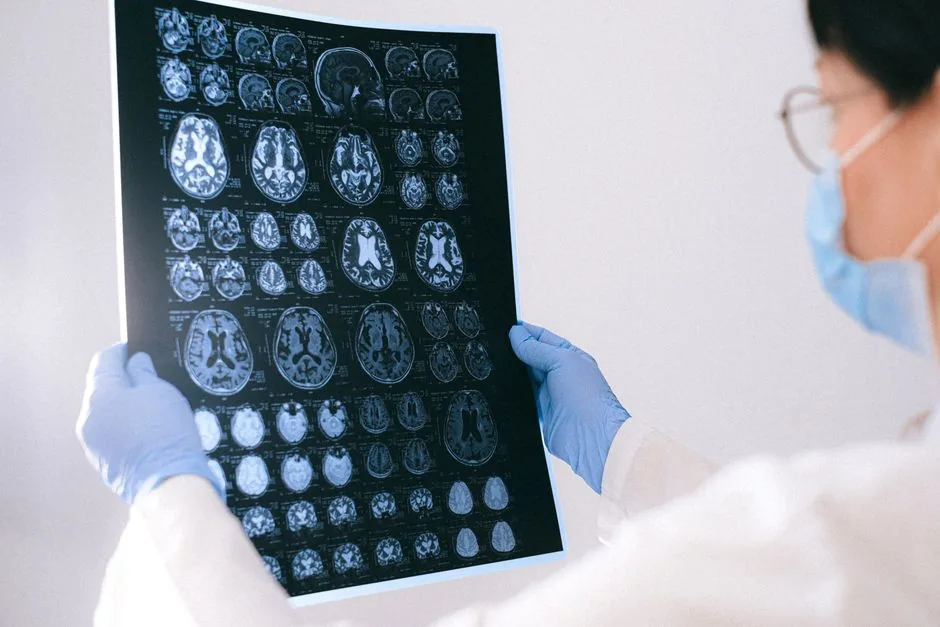
Most studies use a structural MRI, which produces a detailed picture of the brains anatomy. In people with BPD you often see subtle reductions in volume of the frontal lobe, hippocampus, amygdala, orbitofrontal cortex, anterior cingulate and sometimes an increase in the putamen. These differences are tinythink a few millimetresbut theyre consistent enough that researchers can spot a pattern across many scans.
How does a BPD brain differ from a normal brain scan?
| Feature | Typical Brain | BPD Brain |
|---|---|---|
| Frontal Lobe Volume | Standard size for age and gender | Slightly reduced (510% smaller) |
| Amygdala Size | Normal range | Often smaller, indicating emotionprocessing changes |
| Hippocampus | Average volume | Reduced volume linked to stressrelated memory issues |
| Putamen | Typical size | Often enlarged, possibly related to impulse control |
Notice the pattern? The changes are subtle, not dramatic like a stroke or tumor, which is why a scan alone cant hand you a diagnosis.
Is BPD a neurological or psychological condition?
The short answer: both. Your brains wiring (neurology) and life experiences (psychology) dance together. Structural differences give us clues about the neurobiology, but they dont tell the whole storyenvironment, trauma, and coping strategies are equally important.
Can a brain scan diagnose BPD?
No. At present, MRI findings are research tools, not clinical checkboxes. A qualified psychiatrist still relies on interviews, selfreport scales, and a thorough history to make a diagnosis. Think of the scan as a subtle background texturenot the headline.
What are the risks & benefits of getting a brain scan for BPD?
Benefits:Provides objective data that can guide research, help clinicians understand symptom patterns, and sometimes reassure patients that something is visible when they feel dismissed.
Risks:Cost (often a few hundred dollars), waiting time, possible false hope if the scan looks normal, and the emotional impact of seeing a medical image of your own brain.
Its a tradeoff, and the decision should be made with a trusted mentalhealth professional.
Is BPD inherited? Mother vs. father, genetics vs. learning?
Family studies show a modest heritabilityabout 4050% of the risk can be traced to genetics. Both parents contribute, but no single BPD gene exists. Instead, multiple gene variants (e.g.,*5HTTLPR*, *BDNF*) interact with early life stress. So its neither purely genetic nor purely learned; its a complex mashup.
What are the main causes of BPD in females?
Women are diagnosed about three times more often than men. Hormonal fluctuations, higher exposure to certain types of childhood trauma (like emotional abuse), and social expectations around emotional expression all play a role. The brainscan literature hints that some of the structural changes may be more pronounced in females, though research is still catching up.
How do childhood experiences shape the BPD brain?
Early chronic stress can affect the developing limbic system (amygdala, hippocampus). Think of a young tree bent by wind; the shape of the branches (brain pathways) adapts, sometimes leading to heightened emotional reactivity later in life.
What other tests complement a brain scan?
Clinical interviews, the DSM5 criteria checklist, selfreport scales like the Borderline Personality Disorder Severity Index, functional MRI (fMRI) to see activity during emotional tasks, and neuropsychological testing that measures impulse control and memory.
Where can I find reliable BPD imaging studies?
Peerreviewed journals are the gold standard. A recent systematic review summarises the most consistent MRI findings, and the NHS site offers a clear overview of diagnosis and treatment options.
Science Deep Dive
1 Replicated MRI findings
Across dozens of studies, four brain regions repeatedly show volume reductions in BPD:
- Frontal lobe linked to planning and impulse control.
- Bilateral hippocampus memory and stress regulation.
- Amygdala rapid emotional response.
- Orbitofrontal cortex (OFC) decisionmaking and reward evaluation.
Some research also notes an enlarged putamen, which may relate to the compulsive behaviours sometimes seen in BPD.
2 Contested results & why replication matters
Not every study finds the exact same pattern. A few investigations report normal amygdala size, likely due to differences in scanner strength (1.5T vs. 3T), sample size, or the specific emotional tasks participants performed during scanning. This is why metaanalysesstudies that pool data from many labsare crucial for teasing out reliable signals.
3 Functional imaging: beyond structure
Functional MRI (fMRI) looks at blood flow changes while the brain is active. In BPD, fMRI often shows hyperconnectivity of the amygdala during emotional faces, meaning the alarm system stays turned up even when the threat is mild. Another consistent finding: reduced activity in the prefrontal cortex when trying to regulate strong emotions.
4 Genetics meets imaging
Twin studies suggest roughly half of BPD risk is genetic. Certain gene variants (like *5HTTLPR* affecting serotonin transport) have been linked to smaller amygdala volumes, hinting at a pathway from DNA to brain structure to behaviour. A 2023 genomewide association study (GWAS) identified several loci that modestly overlap with cortical thickness findings in BPD.
Supplementary tools
Below is a quick cheatsheet comparing three imaging modalities you might hear about:
| Modality | What It Shows | Typical Use in BPD |
|---|---|---|
| Structural MRI | Graymatter volume, cortical thickness | Research on brainsize differences |
| Functional MRI (fMRI) | Brain activity during tasks | Emotionregulation studies |
| Diffusion Tensor Imaging (DTI) | Whitematter pathways | Connectivity of limbic circuits |
Practical Guide
Assessing personal need
Ask yourself: Do I have neurological symptoms (like seizures or persistent headaches) that need imaging? Am I part of a research study that requires a scan? If youre already in therapy and the focus is on coping skills, a scan may add little practical value.
Finding a qualified provider
Look for a neuroradiology center that has experience with psychiatric imaging. Good questions to ask:
- What Tesla strength does the scanner use?
- Do radiologists specialize in mentalhealth imaging?
- How long will the report take, and will it be explained in plain language?
Costs, insurance, and accessibility
In the U.S., a private MRI can range from $400 to $1,200, while many European health systems cover it if a physician deems it medically necessary. When talking to insurers, frame the request around clinical clarification rather than curiosity. A wellwritten letter from your psychiatrist can make a big difference.
Realworld example
Jenna, a 28yearold graduate student, joined a university research project because she wanted to understand why her intense mood swings felt so biological. The studys MRI showed reduced OFC volume, which her therapist used to tailor dialecticalbehaviortherapy (DBT) modules focused on decisionmaking. Jenna reported feeling validatedshe finally had a concrete image that matched her inner experience.
Common Scenarios
What if my scan looks normal?
Normallooking scans are common. Brain structure is just one piece of the puzzle; neural activity, chemistry, and life history matter just as much. A normal scan doesnt invalidate your strugglesit simply means the current technology isnt sensitive enough to capture every nuance.
Can a brain scan predict treatment response?
Emerging research suggests that people with larger OFC volume may respond better to DBT, while heightened amygdala activation could predict greater benefit from mindfulnessbased therapies. This field is still experimental, so treat such predictions as interesting hints, not hardwired guarantees.
Is a BPD brain scan test coming soon?
Scientists are exploring machinelearning models that combine structural and functional data to flag highrisk patterns. Ethical concernsprivacy, stigma, and the danger of brainbased labelsmean widespread clinical use is still years away.
Do men and women show different brain patterns?
Some studies report stronger amygdala reductions in females, possibly reflecting higher exposure to early interpersonal trauma. However, sample sizes are often small, so the picture remains blurry.
How reliable are online BPD brain scan tools?
Stay away from DIY brainanalysis apps. They lack clinical oversight, use unverified algorithms, and can cause unnecessary anxiety. Always seek interpretation from a licensed neuroradiologist or psychiatrist.
Balancing Reality
Understanding the brain can feel like peeking behind the curtainbut remember, the curtain is only part of the stage. The benefits of neuroimaging are real: they legitimize the lived experience of many, guide research, and sometimes inform treatment tweaks. The riskscost, false hope, and privacy concernsare equally valid.
Bottom line: a BPD brain scan is a valuable research tool and a modest clinical adjunct, not a crystal ball. Use it as part of a broader conversation with your care team, and keep the focus on what helps you feel steadier, more understood, and empowered.
Conclusion
In short, a BPD brain scan can reveal subtle differences in emotionprocessing hubs, yet it isnt a standalone diagnosis. By blending the scans insights with personal history, therapy, and solid clinical judgment, you get a fuller picture of whats happening insideand how to move forward. If youre curious about whether imaging could add value to your journey, talk to your psychiatrist; they can help you weigh the pros, the cons, and the next best steps.