Not medical advice — written by Ngoc Vietsmall, who is not a healthcare professional.
This article summarises publicly available medical information for general readers. It cannot account for your history, medication or symptoms, and it is not a diagnosis or a treatment plan. Any dose mentioned comes from published labelling or guidelines — only your own prescriber can decide what is right for you.
Always speak to a doctor or pharmacist before starting, stopping or changing any treatment. If symptoms are severe or getting worse, seek urgent care. Full disclaimer · How this is written
Imagine youre in the ER and the doctor asks, Is this bleeding or just a blockage? In seconds, a CT scanner can flash a bright white spot that says, Yes, its a hemorrhage. That hyperdense flash is the hallmark of a hemorrhagic stroke CT scan, and its the fastest way doctors know whats happening inside your brain.
In this friendly guide well walk through exactly what that bright spot means, how a hemorrhagic stroke looks on a CT compared to an ischemic stroke, what the images tell doctors about treatment, survival odds, and life after the event. Think of it as a coffeechat with a knowledgeable friendno jargon, just clear, caring answers.
How CT Detects Bleeding Fast
First things first: a noncontrast CT (NCCT) is the goto scan for anyone who might be having a stroke. It takes just a few minutes, and the radiologist can spot fresh blood because blood blocks Xrays more than normal brain tissue. This shows up as a very bright, or hyperdense, areahence the term hyperdense sign.
Why does hyperdensity matter? Fresh blood contains ironrich hemoglobin, which absorbs more Xray photons, making that spot glow white on the monitor. In the first hour after symptoms start, studies report a sensitivity of about 95% for detecting an intracerebral hemorrhage on NCCT (see StatPearls).
Seeing the Difference: Normal vs. Bleed
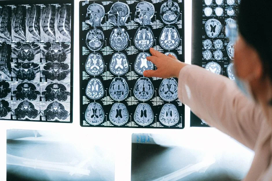
It helps to picture a sidebyside comparison. A normal brain CT is a smooth tapestry of gray and white, with clear spaces for the ventricles and sulci. When a bleed occurs, youll see a distinct, solid white blob that often crowds the surrounding tissue.
Heres a quick checklist you can use if youre ever looking at a scan with a loved one:
- Is there a bright, welldefined area? Thats likely blood.
- Does the shape look round or irregular? Rounded often means a primary bleed; irregular may suggest extension.
- Are the ventricles pushed to one side? Thats a midline shift, a sign the bleed is sizable.
Realworld example: a 58yearold man arrived with sudden rightside weakness. His CT showed a hyperdense spot in the left basal ganglia, confirming an intracerebral hemorrhage and prompting immediate bloodpressure control.
Key CT Features of Acute Hemorrhage
Beyond the bright spot, there are other clues doctors read:
- Surrounding edema: Swelling appears as a hazy, less dense area around the bleed, usually showing up a few hours later.
- Intraventricular extension: Blood can spill into the brains fluidfilled ventricles, which looks like a thin line of density following the ventricles shape.
- Midline shift: The whole brain can be pushed away from the bleed, indicating pressure.
Radiologists often calculate the hematoma volume with the ABC/2 method (length width height 2). This quick estimate helps decide if surgery is needed.
Bleeding vs. Blockage on CT
One of the most common questions is, How can I tell if a stroke is hemorrhagic or ischemic just by looking at the scan? The answer is surprisingly simple:
- Hemorrhagic stroke: Bright white blood, welldefined edges.
- Ischemic stroke: Darker, subtle loss of the graywhite distinction, sometimes called early ischemic changes.
If the CT looks normal but the clinical picture screams stroke, doctors often follow up with an MRI, which can catch early ischemia that CT might miss.
According to the American Heart Association, the gold standard for acute stroke imaging remains the NCCT because it rules out a bleed in under ten minutescrucial when every second counts (AHA).
Types of Hemorrhagic Stroke
| Stroke Type | Typical CT Pattern | Common Location |
|---|---|---|
| Intracerebral hemorrhage (ICH) | Rounded hyperdense mass | Basal ganglia, thalamus, cerebellum |
| Subarachnoid hemorrhage (SAH) | Diffused hyperdensity in sulci & cisterns | Circle of Willis, basal cisterns |
| Intraventricular hemorrhage (IVH) | Blood within ventricles | Lateral ventricles |
| Hemorrhagic transformation | Mixed density, evolving over hours | Often follows an ischemic infarct |
Each type nudges treatment in a slightly different direction. For instance, a pure ICH may be surgically evacuated if its larger than 30mL, while SAH usually calls for endovascular coiling or surgical clipping of an aneurysm.
Survival Rate & Treatment Decisions
Lets face the hard truth: a hemorrhagic stroke carries a higher early mortality than an ischemic one. Metaanalyses show a 30day survival rate ranging from 40% to 70% depending on bleed size, location, and patient age.
But the CT scan is the compass that guides lifesaving moves:
- Bloodpressure control: Rapid reduction (target <140/90mmHg) lowers rebleeding risk.
- Reversal of anticoagulation: VitaminK, prothrombin complex concentrates, or idarucizumab for dabigatran, all guided by how much blood we see.
- Surgical evacuation: If the hematoma is big, causing midline shift, or in an accessible region, neurosurgeons may perform a craniotomy.
- CTA/MRA: Followup CT angiography pinpoints the bleeding sourcean aneurysm, arteriovenous malformation, or hypertensive vessel rupture.
Life After the Scan: Quality of Life
Surviving the bleed is only the first chapter. How you feel weeks, months, and years later depends a lot on what the CT showed. Larger bleeds often mean bigger deficits, but many patients regain independence with rehab.
Studies on quality of life after hemorrhagic stroke reveal that patients who receive early, intensive physical, speech, and occupational therapy report higher satisfaction scoreseven when the initial CT showed a sizable bleed.
Followup CTs are usually scheduled at 24hours, 3days, and 7days. They track hematoma expansion (which can happen in up to 30% of cases) and help decide whether a delayed surgery is needed.
One survivors story: after a lobar bleed in the left temporal lobe, shes now speaking fluently again and enjoys paintingsomething she thought shed lost forever. Her rehab team used the CTs precise location to tailor speech exercises, and the progress was visible on repeat scans.
Putting It All Together
So, what should you walk away with?
- A noncontrast CT can tell a doctor in minutes whether a stroke is bleeding (hyperdense) or blocked (darkening).
- The pattern of the bleedits size, shape, and spreaddirectly influences treatment decisions, from medication to surgery.
- Survival odds improve when the CT findings guide rapid, targeted interventions.
- Even after a big bleed, focused rehabilitation keyed to the CTs map can dramatically boost quality of life.
If youor someone you lovefind yourself facing a hemorrhagic stroke, remember the CT scan is not just a picture; its a roadmap that helps doctors act fast, choose the right treatment, and give you the best shot at recovery.
Next Steps & Resources
Feeling a bit overwhelmed? Thats completely normal. Here are a couple of quick actions you can take:
- Ask your doctor to explain the CT findings in plain languagedont hesitate to ask, What does this bright spot mean for my treatment?
- Look for a certified stroke rehabilitation program near you; many centers tailor therapy based on the exact CT location of the bleed.
- Keep a copy of your scan images. Theyre useful if you ever need a second opinion or want to track progress over time.
Remember, knowledge empowers you to be an active participant in your care. The next time a CT scanner whirs to life, youll know exactly what the bright flash is sayingand youll be ready to act.